The Respiratory System
Functions:
- Provides gas exchange by taking oxygen in and letting carbon dioxide out
- Gives oxygen to blood from the pulmonary artery so that it can be circulated
- Provides gas exchange by taking oxygen in and letting carbon dioxide out
- Gives oxygen to blood from the pulmonary artery so that it can be circulated
Nose
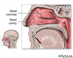
The nose is the main airway of the respiratory system. Anterior nares, a.k.a. nostrils, let air enter the nasal cavity. Cilia in the nasal cavity warm, moisten and clean the air before passing it on through the pharynx. Any foreign debris is trapped by mucus, which is either digested or removed through sneezing and coughing.
Pharynx
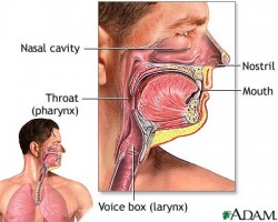
The pharynx (throat) passes air from the nasal cavity onto the larynx. It continues to clean the air as it moves deeper through the respiratory tract.
Larynx
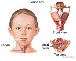
The larynx is commonly called the voice box. It is a shorter tube connected to the pharynx that leads to the trachea. Vocal folds in the larynx contain a narrow vocal ligament. When air pushes upwards from the lungs, these tissues vibrate off of each other to create phonation, or voice sounds.
Trachea
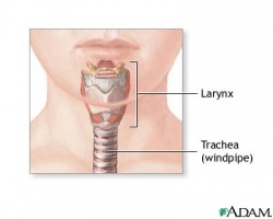
The trachea, also called the windpipe, is a long vertical tube that conducts the passage of air to and from the body. The epiglottis, a flap of tissue located behind the tongue, prevents food from accidently entering the trachea. The inner lining of the trachea produces more mucus to keep removing foreign particles.
Bronchi
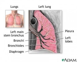
The trachea divides into two smaller tubes called primary bronchi (singular: bronchus). These bronchi lead to either the left or right lung. Each primary bronchi divides into secondary bronchi, and then these divide into tertiary bronchi, which lead to the deep inner lungs.
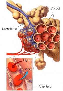
Bronchioles
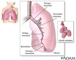
The bronchioles are much smaller tubes made of smooth muscle. The presence of so many tiny brachioles allows for a high surface area for gas exchange to happen so enough oxygen can be received to meet the body's demands.
Lungs
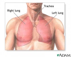
The lungs are the main organs of the respiratory system. They are mainly made up of the complex tissues of the bronchi, bronchioles, and alveolar ducts. The left lung is slightly smaller than the right lung to make room for the heart. The left lung is divided into two lobes (upper and lower), while the right lung is divided into an upper, middle and lower lobe.
Diaphragm
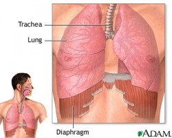
Unlike the heart, the lungs do not contain muscle tissue. Instead, a muscle known as the diaphragm rests underneath the lungs. The diaphragm lifts the ribcage upward to allow the lungs to take in air, then lets the ribcage relax to force air out.
Alveoli

Gas Exchange takes place in alveoli, the 300,000,000 microscopic air sacs located in the lungs. Alveoli are covered in blood vessels so that blood can be quickly oxidized on its way back to the heart.
Images from: http://medlineplus.gov/
